.png)
Severe Tooth Infection Causes Massive Jaw Infection in Young Horse
- Nick Moore DVM
- Apr 1
- 3 min read
A two-year-old horse recently arrived after a 15–17 hour haul from Alabama. The presenting complaint was something we see often: swelling along the jaw and a draining tract. While the case had already undergone some level of workup, the underlying issue had not yet been definitively diagnosed. In equine dentistry, these cases are often more straightforward than they appear. Provided the fundamentals are applied correctly.
There is a tendency in veterinary medicine, especially with complex presentations, to look for rare or unusual causes. But the reality is that most successful outcomes come from doing the basics exceptionally well. Much like elite athletes who master simple movements, good dentistry is built on consistency, pattern recognition, and disciplined diagnostics.
This case followed a very familiar pattern. Facial swelling and purulent drainage strongly suggest dental pathology until proven otherwise. While other causes are possible, infection originating from a tooth is by far the most common explanation.
Upon examination, the degree of mandibular thickening was immediately concerning. The asymmetry between the two sides of the jaw indicated significant bone involvement. The presence of a draining tract confirmed that infection had been present long enough to create a pathway for pressure relief. Radiographs provided clarity. A severe periapical abscess was identified in one of the cheek teeth. Despite previous treatments, the diagnosis at this stage was relatively straightforward: the tooth was non-viable and required extraction.
This highlights a critical point—treatment without diagnosis is often ineffective. Flushing, antibiotics, and symptomatic care cannot resolve an abscess when the source of infection remains in place. In this case, the infected tooth had essentially become a foreign body. As with any foreign body infection, removal is mandatory for resolution.
Further examination revealed that the problem extended beyond a single tooth. Chronic infection had progressed into the surrounding bone, resulting in osteomyelitis and the formation of necrotic bone fragments (sequestra). This significantly increased the complexity of the case. Not because the extraction itself was difficult, but because complete resolution required removal of both the diseased teeth and the compromised bone.
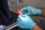
The procedure was performed standing, using appropriate sedation and regional anesthesia.
Avoiding general anesthesia reduces risk, lowers cost, and improves recovery. Particularly when the team is experienced, and the process is well-structured.
Once the affected teeth and necrotic bone were removed, improvement was rapid. Within 24 hours, drainage had significantly decreased. Within days, external tracts began to close. This dramatic turnaround reinforces how much pain these horses endure and how quickly they recover when the source is addressed.
Interestingly, this horse presented with multiple affected teeth, which is uncommon at such a young age. It serves as a reminder that while patterns are helpful, clinicians must always evaluate the entire mouth and remain open to atypical presentations.
Ultimately, the success of this case came down to a systematic approach: accurate diagnosis, logical sequencing, and efficient execution. These are not advanced concepts, but they are often overlooked.
For horse owners, the message is simple: swelling, drainage, or unexplained discomfort should never be ignored.
For veterinarians, the takeaway is just as clear: mastery of the fundamentals is what makes complex cases manageable.
Want to Learn More?
If you're a veterinary professional interested in expanding your knowledge in equine dentistry, we offer training courses on equilibration, diagnosis, endoscopic assessment, and more. You can always contact us at mooredvmeducation@gmail.com or call the office at 512-508-8141.